DNasing RNA, Round II-V
DNasing, Rounds II-V
I performed 3 rounds of DNase reactions (n=30 per round) on the Oly larval and ctenidia RNA. I followed the same protocol as Round 1, using 50 uL RNA for each sample, 5 uL buffer, 1 uL DNase, and 5 uL inactivation reagent.
There were two samples - 38 and 46 (extracted previously in 2018) - which had less than 50 uL total volume in the original RNA sample. For these I used 25 uL RNA, 2.5 uL buffer, 1 uL DNase and 2.5 uL inactivation reagent.
Several of the samples also required dilution prior to DNasing, since the maximum amount of RNA in a 50 uL reaction is 10 ug. For these I added 50 uL DEPC-treated water and vortexed to mix, prior to DNasing: 477, 43, 564, 291, 304, 306, 309, 315, 316, 317, 321, 325, 335, 345.
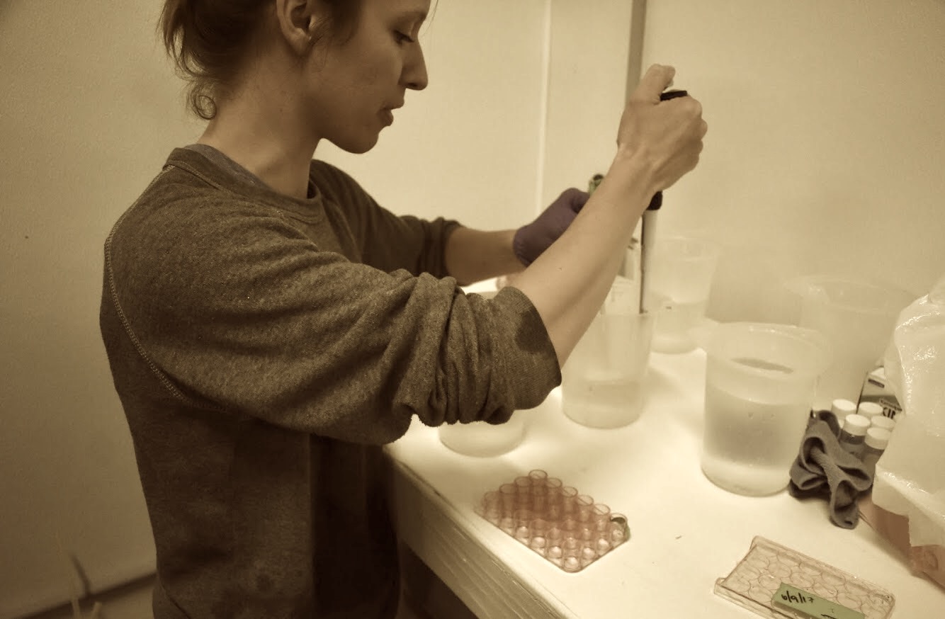
Some images of the Turbo DNase process
This is the Turbo DNase kit I used.

In it is: A) Buffer B) TurboDNase enzyme C) Inactivation Reagent D) Ultrapure water (to resuspend inactivation reagent, if it has dried).
NOTE: the kit says it’s good for 50 reactions, but that’s for 100 uL volume / reaction. For me, the limiting factor was the DNase enzyme - there was 120 uL DNsae (used 1 uL per sample) - so I did ~110 samples with one kit before pulling DNase from the other.

2 sets of labeled 0.5 mL tubes. Step 1 is to transfer 50 uL RNA into one set of tubes.
After adding buffer (10% of RNA volume, in my case 5 uL), and DNase (1 uL), I vortexted gently and incubated at 37C for ~25 minutes in the Thermo Cycler. It takes no time to heat up, keeps track of the time. Maximum # tubes at once is 30.


I forgot to get images of the inactivation reagent step - the solution is milky white, and after mixing and incubating at rooom temp for 5 minutes, samples are centrifuged to “pellet” the reagent, then the supernatant containing RNA is transferred to the 2nd batch of tubes. NOTE: I held everything on ice while working.
qPCR to check for DNA contamination
I ran 3 more batches of qPCR to check for DNA contamination.
For these batches, I selected Olympia oyster primers ordered by Jake


Primers are located in the freezer in FTR 213.

Found the Oly primers using the database.

I created working stocks of my primers - diluted 12 uL each primer in 108 uL DECP-treated water, creating 120 uL at 10 uM for each.
Created a mastermix for all reactions using the following calculations, then pipetted 19 uL mastermix onto qPCR plates, and 1 uL each sample in duplicate.
| Date | 1 plate , 32 rxns | All 3 reactions |
|---|---|---|
| # Samples | 32 | 96.0 |
| # Reactions | 68 | 204.0 |
| Template (RNA sample) (uL) | 68.0 | 204.0 |
| Sso Fast (uL) | 748.0 | 2,244.0 |
| Pf, 10 uM (uL) | 37.4 | 112.2 |
| Pr, 10 uM (uL) | 37.4 | 112.2 |
| DEPC-treated water (uL) | 598.4 | 1,795.2 |
| Total Volume in mastermix (uL) | 1,421.2 | 4,263.6 |
Round II
Results look good - only the positive control (DNA sample 1A, in green) had a detected melt temperature. I realized later that I accidentally used the wrong qPCR protocol - I used “CFX_2StepAmp60_EVAGreen+Melt.prcl” instead of “CFX_2StepAmp_EVAGreen+Melt.prcl”. The difference is that step 3 is 60C, while in the correct protocol step 3 is 55C. For my purposes, and since results look good, it seems that this error does not warrant a re-do. I’ll check with the lab, though.

Round III
Results look a bit weird, different than before. The fluorescence (RFU) goes up at high temperatures - not sure if this indicates an issue. Also, two samples had melt temperatures - 543 and 475 - but only in 1 rep for each.


Round IV
Results look okay, except for one well again had a weird increase in RFU at high temps (sample 476, rep 2). However, there was no melt temperature measured in the RNA samples.

All edited CFX files (with sample names, colors) are located in the qPCR-DNA-contamination directory in my O.lurida_Stress repo.
Raw CFX files are saved to owl here